Bioanalytik
Our platform specializes in offering a range of techniques for biomolecular interaction analysis and is specifically designed to facilitate academic collaborations.

Our platform specializes in offering a range of techniques for biomolecular interaction analysis and is specifically designed to facilitate academic collaborations.

We provide training on the following instruments and offer assistance in data interpretation: MicroCal PEAQ-ITC for isothermal calorimetry (ITC), NanoTemper Monolith NT.115 (Microscale thermophoresis [MST]), NanoTemper Prometheus NT.48 (nanoDSF), FluoroMax3 (fluorimetry), DynaPro NanoStar Wyatt Technologies (DLS), and Varian260 (AAS). After receiving an introduction, users are able to operate these instruments themselves, and there is a fixed fee per hour for their usage. Upon request, we can establish scientific collaborations involving these instruments.
For more advanced techniques such as SPR (Surface Plasmon Resonance), which users are not be able to perform themselves, we offer a full-price service or a scientific collaboration, providing support in technical and scientific matters related to your project. Collaborative projects include consultation, project planning, and data evaluation.
We also offer measurement services to other academic institutions in the Munich area upon request. In such cases, project costs and user fees will be allocated according to a cost plan.
Please take note of our user regulations and protein QC guidelines.
For further information, questions, and support, please contactbioanalytics laboratory rooms G02.037/G02.039
Please take note of our user regulations and protein QC guidelines. Login for availabilit check and booking (Fluorimeter, AAS, DLS/MALS and Thermophoresis)
Interactions between biomolecules play a crucial role in various cellular processes. These interactions can involve protein-protein, DNA-protein, lipid-protein, or protein-low molecular weight ligand interactions. To understand the functioning of proteins, nucleic acids, lipids, and other molecules in a biological system, it is essential to identify their interaction partners and characterize their interactions. The characterization of biomolecular interactions requires knowledge of affinity, the number of binding sites, and thermodynamic properties.
By performing a simple ITC experiment, it is possible to determine parameters such as binding affinity (KD), stoichiometry, enthalpy (ΔH), and entropy (ΔS). Thermodynamic parameters such as ΔH and ΔS provide valuable insights into complex formation and possible conformational changes of a binding partner after the binding event.
The Bioanalytic Core Facility offers the usage of the PEAQ-ITC microcalorimeter from Malvern. For more details about our PEAQ-ITC instrument, please visit the MalvernPanalytical
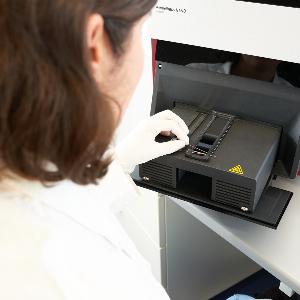
Mobile particles exhibit different responses to the force of a temperature gradient. This ohenomenon is called thermophoresis. The Bioanalytics core facility is equipped with a Monolith NT.115 device (NanoTemper) Microscale-Thermophoresis device that is based on detection of fluorescent proteins or dyes.
Therefore, affinities of biomolecular interactions of dye-labelled proteins or DNA e.g. with any non-labelled compound and can easily be measured in solution in any buffer or complex bioliquid, using only a tiny amount of sample. We offer training at the instrument and help interpreting the data. Usage is at cost, so users only have to pay consumables (glass capillaries) that have to be bought by themselves.
Please use the Nanotemper online shop to purchase consumables
Biacore systems define the characteristics of proteins in terms of their specificity of interaction with other molecules, the rates at which they interact (binding and dissociation), and their affinity (how tightly they bind to another molecule). Binding can be monitored in real-time in a labelfree system. We use the latest technology for measurements on the Biacore T200 system. We support your project in technical and scientific questions.
Bei allen zellulären Prozessen spielen Wechselwirkungen zwischen Biomolekülen eine zentrale Rolle. Dies können z.B. Interaktionen zwischen Protein-Protein, DNA-Protein, Lipid-Protein, oder Protein-niedermolekularer Ligand sein. Wenn man die Funktionsweise von Proteinen, Nukleinsäuren, Lipiden oder anderen Molekülen in einem biologischen System verstehen will, ist es essentiell deren Interaktionspartner zu kennen sowie deren Wechselwirkung zu charakterisieren.
Die Charakterisierung von biomolekularen Interaktionen erfordert Kenntnisse über die Affinität, die Anzahl der Bindestellen sowie die thermodynamischen Eigenschaften. Durch ein einfaches ITC Experiment ist es möglich, Parameter wie Bindeaffinität (KD), Stöchiometrie, Enthalpie (ΔH) und Entropie (ΔS) zu bestimmen.
Die Kenntnis thermodymischer Parameter wie ΔH und ΔS gibt weitere Aufschlüsse über die Komplexbildung, aber auch über mögliche Konformationsänderungen eines Bindepartners nach dem Bindeereignis. Dies ist mit dem PEAQ-ITC Mikrokalorimeter der Firma Malvern möglich; das Gerät befindet sich in der Serviceeinheit Bioanalytik.
Die Charakterisierung isolierter Biomoleküle ist ein wesentlicher Bestandteil von molekularbiologischen Forschungsstudien. Um beispielsweise die Funktionsweise von Proteinen zu verstehen, ist es notwendig, deren exakte biophysikalische Eigenschaften zu kennen.
Durch die Kombination von MALS ("Multiple Angle Light Scattering") und DLS ("Dynamic Light Scattering") können die absolute molare Masse von Biomolekülen sowie auch deren Radii exakt und kalibrationsfrei bestimmt werden, so dass klare Aussagen über den Oligomerisationszustand und die Konformation von Makromolekülen, wie Proteinen, anderen natürlichen Biopolymeren oder Nanopartikeln, getroffen werden können.
Diese Kenntnis lässt aber Rückschlüsse über Aggregation, Stabilität und Homogenität von Biomolekülen zu, und ist daher zur optimalen Vorbereitung von Proben für Folgestudien wie z.B. biomolekulare Interaktionsstudien oder Kristallisation äußerst wichtig. Die Serviceeinheit Bioanalytik ist mit einem DLS-Detektor (Wyatt DynaPro Nanostar) und einem MALS-Detektor (Wyatt miniDAWN TREOS) ausgestattet. Die Kombination beider Techniken bietet eine äußerst sensitive Messmethode, um die oben genannten biophysikalischen Eigenschaften von z.B. Proteinen zu bestimmen.
Atomic Absorption Spectroscopy (AAS) uses the absorption of light to measure the concentration of gas-phase atoms. Since samples are usually liquids or solids, the analyte atoms or ions must be vaporized in a flame or graphite furnace. The atoms absorb ultraviolet or visible light and make transitions to higher electronic energy levels. The analyte concentration is determined from the amount of absorption.
Applying the Beer-Lambert law directly in AA spectroscopy is difficult due to variations in the atomization efficiency from the sample matrix, and nonuniformity of concentration and path length of analyte atoms (in graphite furnace AA). Concentration measurements are usually determined from a working curve after calibrating the instrument with standards of known concentration.
The light source is usually a hollow-cathode lamp of the element that is being measured.
To date we have the following lamps:
We offer training at the instrument and help interpreting the data. Usage is at cost, so users only have to pay consumables (gas/standards, payed by usuage per hour) and a maintainance fee (fixed fee payed per measurement).
Fluorescence spectroscopy is a type of electromagnetic spectroscopy which analyzes fluorescence from a sample. It involves using a beam of light, usually ultraviolet light, that excites the electrons in molecules of certain compounds and causes them to emit light of a lower energy, typically, but not necessarily, visible light.
Conformational changes of a protein (e.g. by binding a ligand) can be investigated changes in the fluorescence spectrum. The fluorescence of a folded protein is a mixture of the fluorescence from individual aromatic residues. Most of the intrinsic fluorescence emissions of a folded protein are due to excitation of tryptophan residues, with some emissions due to tyrosine and phenylalanine; but disulfide bonds also have appreciable absorption in this wavelength range.
Typically, tryptophan has a wavelength of maximum absorption of 280 nm and an emission peak that is solvatochromic, ranging from ca. 300 to 350 nm depending in the polarity of the local environment. Hence, protein fluorescence may be used as a diagnostic of the conformational state of a protein.
Furthermore, tryptophan fluorescence is strongly influenced by the proximity of other residues (i.e., nearby protonated groups such as Asp or Glu can cause quenching of Trp fluorescence). Also, energy transfer between tryptophan and the other fluorescent amino acids is possible, which would affect the analysis, especially in cases where the Förster acidic approach is taken. In addition, tryptophan is a relatively rare amino acid; many proteins contain only one or a few tryptophan residues.
Therefore, tryptophan fluorescence can be a very sensitive measurement of the conformational state of individual tryptophan residues. The advantage compared to extrinsic probes is that the protein itself is not changed. The use of intrinsic fluorescence for the study of protein conformation is in practice limited to cases with few (or perhaps only one) tryptophan residues, since each experiences a different local environment, which gives rise to different emission spectra.
We offer training at the instrument and help interpreting the data. Usage is at cost, so users only have to pay a maintainance fee (fixed fee payed per measurement).